When you are down with influenza, your hunger, thirst and energy, all go down. But have you ever wondered how your brain senses the infection? And orders your body to rest? Who are the messengers who give your brain this information?
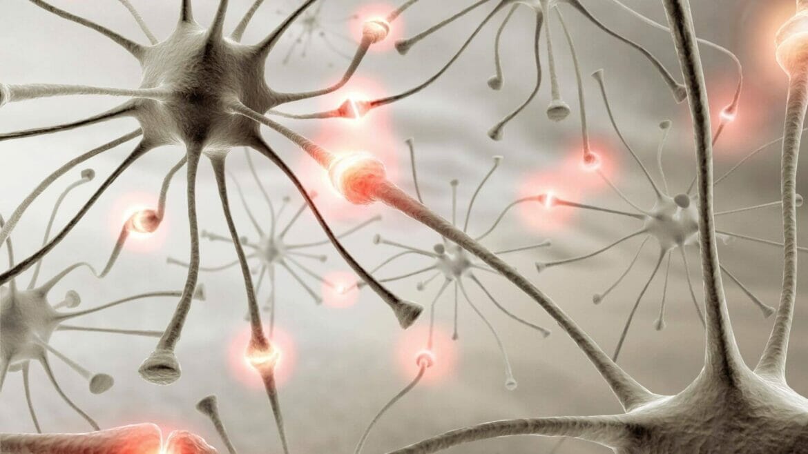
In a paper published in Nature, scientists identified neurons in mice sending signals to the brain, triggering a decrease in movement, hunger and thirst. The neurons located in the throat sensed the influenza infection and notified the brain.
Further, the scientists say similar neurons in other parts of the body may notify the brain about different infections.
A paradigm shift to the previous understanding
This study is a paradigm shift in understanding what notifies the brain about an infection.
The study’s co-author Stephen Liberles, a neuroscientist at the Harvard Medical School in Boston, says that previously, it was unclear how the brain became aware of different infections.
Generally, scientists believe that messenger molecules at the infection site travelled in the bloodstream to notify the brain of the infection, which kickstarts the sickness-behaviour program.
The scientists thought Prostaglandins made in the infected tissues were the signalling chemicals. This was believed because Aspirin and Ibuprofen significantly suppressed sickness behaviours, including pain and inflammation. Both medicines work by inhibiting an enzyme that produces prostaglandins. Hence the scientists thought that prostaglandins relay the information about the infection to the brain.
The new findings
EP3 is the specific prostaglandin receptor thought to notify the brain about infection and generate sickness behaviours. This receptor is present in the brain as well.
During the study in mice, the scientists deleted the brain’s EP3 receptors and infected the mice with a flu virus. The animals still changed their behaviour, implying that the blood-borne prostaglandins are not the infection messengers to the brain.
The scientists also found the key agents to be specific EP3-containing neurons in the mice’s neck area. These neurons have branches assumed to be like a dedicated highway to the brain, enabling effective and accurate transmission.
So, the new narrative of flu illness is:
- The Flu virus enters the airway, infecting throat cells
- Triggers Prostaglandin production in the throat, causing inflammation
- Neurons in the throat area sense prostaglandin production and hence the infection. Infection alert travels along the neurons’ branches on a dedicated highway to the brain
Neurons on dedicated pathways
The study’s authors noted that information relayed through such dedicated neural networks could give the brain information about where exactly the infection is occurring.
They also assumed that many other types of neurons would have receptors for prostaglandin and other immune-related signals. And so, there will be neural pathways dedicated to information transfer to the brain, including ones for gut infections.
What does this development tell us?
This study finds that neurons in the throat and tonsils relay information about an upper respiratory flu infection to the brain. It also assumes as the infection moves from the upper respiratory tract to the lower respiratory tract, the neurons transmitting the information and triggering sickness behaviours will not be the ones in the throat area but different. Presumably, such dedicated neural pathways are present throughout the body to sense prostaglandin and other immune-related signals, notifying the brain about the infection and the place infected. In response, the brain triggers sickness behaviours such as reduced hunger and movement in the case of a flu infection.
Images: canva.com
